
The reconstruction of the anterior segment demands high cervicosurface adaptation, absolute field decontamination, and precise control over polymer layer thickness to minimize polymerization vectors pulling away from fragile enamel margins.
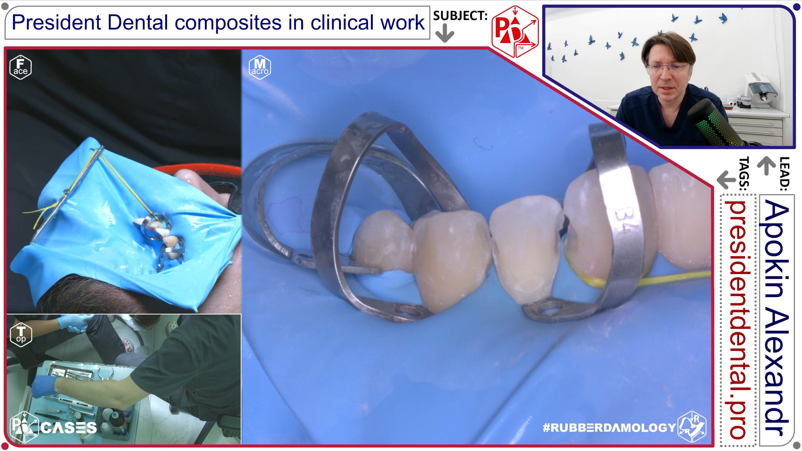
Restoration cervical area tooth 21, 22, 23 caries media with retractors B4, Prebond, Stark bulkfill, Dynamic Plus | President Dental Case 0001 ENG 4K
Case 0001: Multi-Unit Cervical Defect Management in the Maxillary Esthetic Zone (Teeth 21, 22, 23)
- Primary Clinical Pathology: Extensive Class V cervical carious degradation (caries media) crossing the cementoenamel junction (CEJ), exhibiting highly sclerotic dentin borders.
- Isolation and Soft-Tissue Retraction Matrix: Absolute field isolation via a heavy latex sheet. Deep mechanical tissue deflection executed using independent Brinker B4 gingival retractors stabilized under high lateral frame tension to fully reveal the subgingival cavosurface margins.
- Biomimetic Material Architecture: Chemical surface conditioning via 5th-generation total-etch Prebond adhesive. Structural core base built using Stark Bulkfill to establish a low-shrinkage dentin core, overlaid with Dynamic Plus micro-hybrid universal composite to replicate the high-stiffness facial enamel shell.
- Clinical Execution Strategy: Micro-kinetic alumina sandblasting (Al2O3, 50μm) was used to break down the acid-resistant aprismatic enamel and clean the subgingival margins. The Stark Bulkfill base was placed in a single 3.5 mm increment to eliminate internal voids, followed by anatomical sculpting of the Dynamic Plus layer using a fine composite brush to match the native horizontal macro-texture of the incisor crowns.
Restoration cervical area tooth 21, 22, 23 caries media with retractors B4, Prebond, Stark bulkfill, Dynamic Plus | President Dental Case 0001 ENG 4K
1. Initial Clinical Status and Defect Topography
The patient presented with multiple carious lesions localized in the cervical third of the maxillary left central incisor, lateral incisor, and canine (teeth 21, 22, and 23 using the FDI world dental numbering system). Clinical and visual assessment confirmed mid-stage dental caries (caries media) compromising the gingival margins.
The defects extended close to the cementoenamel junction (CEJ), showing variable depths and structurally compromised enamel margins. Because these lesions were located within the aesthetic zone, the clinical objective was twofold: achieving absolute marginal adaptation to eliminate the risk of secondary caries, and restoring natural optical properties without visible transition lines.
2. Isolation and Soft-Tissue Management: The #RSBC Protocol


Managing multiple cervical defects simultaneously requires profound control over the gingival architecture to expose subgingival margins and completely prevent moisture contamination:
- Rubber Dam Matrix: A heavy-gauge latex dam was selected to exert steady peripheral retraction. Individual perforations were punched precisely to handle the anatomical transition from the central incisor to the prominent canine bulge.
- Mechanical Retraction via B4 Clamps: To achieve complete visual access below the free gingival margin without surgical gingivectomy, specialized B4 cervical retractors were stabilized on each target tooth. The apical adaptation of the B4 clamp safely displaces the dam and soft tissue gingivally, exposing the entire subgingival boundary of the lesion.
- Marginal Sealing: The sulcular environment was hermetically sealed against moisture, blood, and crevicular fluid by combining the rubber dam inversion with a dedicated liquid dam matrix applied around the active jaw elements of the B4 clamps.
3. Substrate Conditioning and Adhesive Protocol
To ensure long-term retention of the composite material against cervical flexure forces (abfraction-like stress), the substrate was conditioned according to strict #RSBC parameters:
[Micro-Air Abrasion via Al2O3 (27–50 μm)]
                       │
                       ▼
     [Selective Enamel Etching (37% H3PO4)]
                       │
                       ▼
[Active Hybridization via Pre Bond (President Dental)]
- Micro-Abrasive Preparation: The sclerotic and altered dentin surfaces within the cervical lesions were treated using an intraoral air-abrasion unit with aluminum oxide ($Al_2O_3$) particles (27–50 μm) at a stable pressure of 2 bar. This eliminates the highly organized aprismatic layer and maximizes the surface free energy of the dentin substrate.
- Acid Etching: A selective enamel-etching technique was carried out using 37% phosphoric acid ($H_3PO_4$) gel. The enamel margins were etched for 15 seconds, ensuring the gel did not cause dentinal hypersensitivity or over-etching of the deep cervical walls. The area was rinsed thoroughly with a water-air spray for 20 seconds and gently air-dried.
- Adhesive Application: Pre Bond (President Dental) universal adhesive was actively massaged into the exposed dentin for 20 seconds to facilitate monomer hybrid layer formation. The solvent was carefully evaporated using a gentle, dry air stream until a motionless, glossy adhesive layer was confirmed, followed by a 20-second light-polymerization cycle.
4. Biomimetic Reconstruction and Stratification Strategy
The restoration protocol utilized a dual-material workflow designed to neutralize polymerization shrinkage stress while maintaining anterior aesthetics:
- Stress-Absorbing Base Layer: To ensure void-free marginal adaptation along the sharp internal line angles of the cervical floor, Stark (Bulk Fill Flowable) was applied as an adaptive initial layer. Its flowable adaptation provides stress-buffering capacity, counteracting the high C-factor inherent to Class V configurations.
- Anatomical Shading and Contour: The final outer layers of teeth 21, 22, and 23 were reconstructed using Dynamic Plus universal microhybrid composite. Its non-sticky handling and high filler load allowed for precise anatomical sculpting right up to the displaced CEJ line. The microhybrid formulation provides excellent polishability and refractive properties, seamlessly blending the restorative margin with the natural tooth structure.
- Finishing & Polishing: Following incremental light-curing, initial contouring was performed while the B4 clamps maintained soft-tissue retraction, ensuring no overhangs were left to cause plaque accumulation post-operatively.
CONCLUSIONS
- Efficiency of B4 Retractors: The concurrent restoration of teeth 21, 22, and 23 demonstrates that B4 cervical retractors are vital for multiple Class V operations. They guarantee steady tissue displacement and stable visibility without requiring separate isolation setups for each unit.
- Optimized Marginal Integrity: Combining a flowable base (Stark Bulk Fill) with a high-density universal composite (Dynamic Plus) creates an ideal biomechanical match for cervical restorations. The flowable base minimizes marginal stress, while the microhybrid exterior delivers the wear resistance and aesthetic finish required for the anterior segment.
- Adhesive Reliability via Pre Bond: Micro-abrasion paired with the active application of Pre Bond ensures reliable long-term bond strengths, reducing the clinical failure rates typically associated with cervical margins under occlusal loading and structural flexure.
Composite restoration tooth 13 Prebond, Dynamic Plus | President Dental Case 0009 ENG 4K
Case 0009: Monolithic Class III/V Transitional Restoration of the Maxillary Canine (Tooth 13)
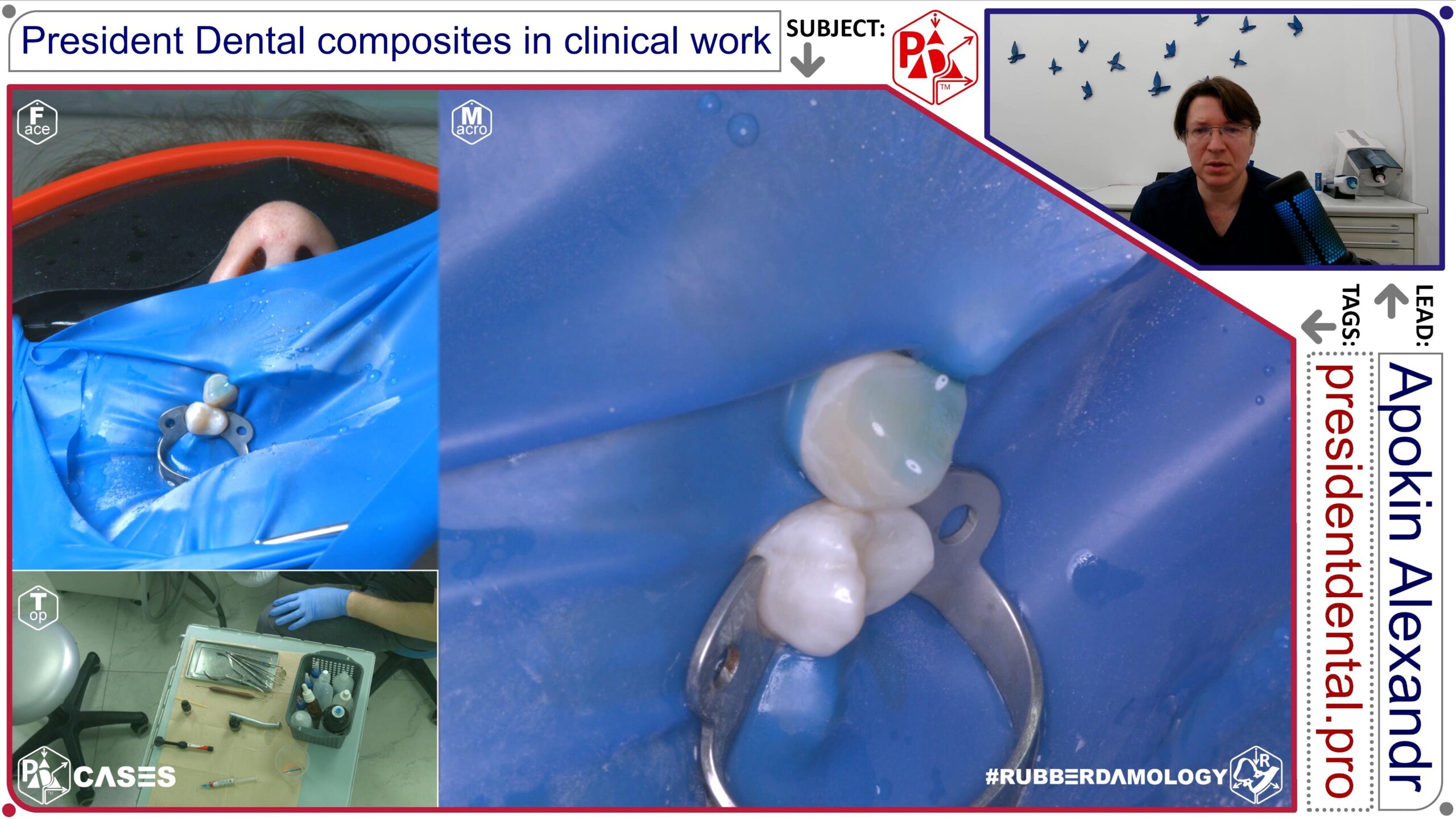
- Primary Clinical Pathology: Isolated, deep disto-cervical Class III cavity configuration merging into a structural Class V erosion defect (caries media) on the prominent root bulge of the upper right canine.
- Isolation and Soft-Tissue Retraction Matrix: Heavy-gauge rubber dam isolation anchored distally on tooth 16. Localized tissue displacement achieved by adapting a cervical clamp to expose the subgingival margins without encroaching on the interproximal embrasure space.
- Biomimetic Material Architecture: Prebond total-etch adhesive network paired with a monolithic layering approach using Dynamic Plus micro-hybrid universal composite.
- Clinical Execution Strategy: After removing the carious tissue, the enamel margins were prepared with a long, regular bevel to ensure a smooth aesthetic transition. The highly filled Dynamic Plus composite was packed against the internal line angles to counteract the high C-factor of the deep cervical box, ensuring tight adaptation along the internal margins.
This clinical case report details the step-by-step direct aesthetic restoration of the vestibular surface of tooth 13 (upper right canine). Conducted by Dr. Alexander Apokin as part of the President Dental demonstration curriculum, this case highlights a strictly minimally invasive approach executed under the paradigm of the international RSBC protocol (Rubberdam – Sandblasting – Bonding – Composite).
1. Initial Clinical Status and Defect Topography
- Patient Presentation: The patient presented with a superficial structural defect/discoloration localized entirely on the vestibular (facial) aspect of tooth 13.
- Pre-operative Management: Local infiltration anesthesia was administered in the mucobuccal fold adjacent to the canine to ensure patient comfort during tissue retraction and conditioning.
- Conservative Strategy: A key feature of this case is the complete elimination of rotary instruments. No diamond or carbide burs were introduced. The treatment plan relied solely on chemical and kinetic surface conditioning to maximize the preservation of healthy enamel rods.
2. Isolation and Soft-Tissue Management: The #RSBC Protocol
Absolute moisture control is the foundational pillar of the RSBC concept, ensuring the long-term integrity of the adhesive interface.
- Barrier Selection: Complete isolation of the working zone was achieved using a heavy-gauge latex rubber dam sheet (Sanctuary Dental). The heavy-gauge material provides optimal elasticity and superior tissue retraction.
- Clamp Anchorage: A reliable double-bowed retention clamp (TOR VM, generic equivalent to the U2 style) was stabilized distally on the premolar region (tooth 14).
- Inversion and Retraction: The rubber dam was inverted sulcularly around the cervical margin of tooth 13. This isolates the vestibular enamel from sulcular fluid (crevicular seepage), saliva, and moisture from the patient’s breath, while simultaneously safeguarding the patient’s airway from micro-particles during the subsequent sandblasting stage.
3. Substrate Conditioning and Adhesive Protocol
The adhesive interface was optimized through a synergistic mechanical and chemical conditioning sequence:
- Kinetic Micro-Abrasion (Sandblasting): The vestibular enamel surface was conditioned using an air-abrasion handpiece loaded with fine aluminum oxide ($\text{Al}_2\text{O}_3$) particles. This process strips away the organic biofilm, removes extrinsic stains, and micro-textures the superficial enamel layer to drastically increase the surface area available for chemical bonding.
- Total-Etch Technique: A phosphoric acid etching gel (President Dental) was applied across the entire sandblasted enamel zone. Upon contact, gas micro-bubbles formed organically, signaling the dissolution of the smear layer. The gel was actively agitated with a microbrush to optimize penetration. This total-etch step is clinically critical, as the acid dissolves and flushes out any microscopic $\text{Al}_2\text{O}_3$ granules wedged within the enamel prisms.
- Evaporation and Bonding: The acid gel was thoroughly rinsed with a copious water-air spray. The substrate was air-dried until the enamel displayed a uniform, chalky-white matte appearance. Next, Prebond (President Dental), a high-stability 5th-generation single-component adhesive, was dispensed directly via its translucent, fine-tip dropper bottle. The adhesive was gently air-thinned to form a homogeneous micro-layer and light-cured for 3 seconds at maximum intensity.
4. Biomimetic Reconstruction and Stratification Strategy
The final phase focused on anatomical replication and precise management of the composite material’s physical handling properties:
- Material Selection: The vestibular morphology was restored using Dynamic Plus (President Dental), a universal microhybrid composite known for its wear resistance and structural predictability, selected in shade A3 to match the patient’s natural dentin/enamel chroma.
- The “Glove-Drop” Modeling Technique: To facilitate smooth adaptation of the dense microhybrid paste, the operator placed a small droplet of modeling resin on the back of their left-hand treatment glove. The tip of the manual modeling instrument (composite spatula) was lightly wiped against this resin before contacting the composite. This creates a micro-thin lubricating barrier on the instrument, preventing the composite from sticking to the metal or pulling away from the delicate enamel margins during sculpting.
- Anatomical Sculpting: The composite was adapted incrementally to rebuild the organic facial contours, proximal transitions, and emergence profile of the canine. The material was sculpted smoothly to blend seamlessly into the surrounding tooth structure, followed by final multi-directional photo-polymerization.
CONCLUSIONS
- Synergy of the RSBC Workflow: The strict execution of the “Rubberdam – Sandblasting – Bonding – Composite” sequence proves that even simple vestibular defects yield highly predictable, long-lasting aesthetic results when standard protocols are maintained without compromise.
- Ultra-Conservative Dentistry: By substituting traditional burs with aluminum oxide air-abrasion, the operator achieved maximum enamel preservation, proving that superficial defects can be thoroughly conditioned without destructive macro-preparation.
- Routine Isolation Efficiency: Implementing a rubber dam should not be seen as a time-consuming luxury. With appropriate clamp selection and a streamlined approach, it takes minimal clinical time while serving as the primary guarantor against micro-leakage and adhesive failure.
- Optimized Composite Handling: Utilizing a trace amount of modeling resin on hand instruments significantly improves the adaptability of microhybrid materials like Dynamic Plus. This technique eliminates structural voids and surface defects, facilitating a seamless transition from synthetic biomaterial to natural tooth structure.
Composite restoration of tooth 11, 21 with sylicone key Prebond, Stark bulkfill, Dynamic Plus, Flow | President Dental Case 0013 ENG 4K
Case 0013: Index-Guided Multi-Unit Structural Reconstruction of Maxillary Central Incisors (Teeth 11, 21)
- Primary Clinical Pathology: Extensive Class IV coronal fractures involving substantial loss of palatal enamel, internal dentin structures, and incisal edge perimeters.
- Isolation and Soft-Tissue Retraction Matrix: Absolute quadrant isolation spanning from tooth 14 to 24, utilizing inversion ligatures to keep the interdental papillae clear of the bonding zone.
- Biomimetic Material Architecture: Prebond total-etch adhesive, Stark Bulkfill for internal dentin replacement, Dynamic Plus micro-hybrid layers for facial/incisal enamel reconstruction, and President Dental Flow to optimize internal adaptation.
- Clinical Execution Strategy: A preoperative silicone key index, fabricated from a diagnostic wax-up, was positioned to guide the palatal shell reconstruction. A micro-thin (0.5 mm) wall of Dynamic Plus was layered directly onto the silicone matrix to form the palatal enamel backing. Stark Bulkfill was then used to replace the missing dentin volume within the core of the fractured zone, leaving precise space for incisal halo characterization before finalizing the facial layer with Dynamic Plus.
Case 0037: Comprehensive Mandibular Labial Cervical Restoration Block (Teeth 41, 42, 43, 44, 45, 46)
- Primary Clinical Pathology: Linear, widespread cervical carious lesions (caries media) extending across six contiguous teeth in the lower right quadrant, accompanied by gingival recession.
- Isolation and Soft-Tissue Retraction Matrix: Total mandibular isolation anchored bilaterally. Simultaneous soft-tissue retraction managed by stacking Brinker B1 and Brinker B4 retractors sequentially across the quadrant, stabilized by NovaTwist accessory loops to prevent clamp rotation.
- Biomimetic Material Architecture: Prebond universal total-etch adhesive, single-shade optical adaptation via ZenChroma, backed by Stark Bulkfill bases in deeper posterior box extensions.
- Clinical Execution Strategy: Aluminum oxide sandblasting was executed across all six cervical fields to clear organic debris from the gingival sulcus. Prebond was applied and thoroughly light-cured across the entire block. ZenChroma single-shade composite was then systematically placed across the anterior segments, utilizing its omni-chromatic blending properties to naturally mirror the diverse tooth shades across the canine-premolar transition zone.
Case 0047: Multi-Unit Interproximal Anterior Reconstruction using Sectional Matrices (Teeth 13, 12, 11, 21, 22)
- Primary Clinical Pathology: Extensive, mirror-image Class III interproximal carious lesions (caries media) undermining the marginal ridges and proximal contact networks of the maxillary anterior segment.
- Isolation and Soft-Tissue Retraction Matrix: Upper arch rubber dam isolation from tooth 15 to 25. Interdental spaces managed using ultra-thin anterior TOR VM sectional matrix foils.
- Biomimetic Material Architecture: Prebond total-etch adhesive network, Stark Bulkfill for proximal box structural bases, and Dynamic Plus micro-hybrid composite for surface aesthetics.
- Clinical Execution Strategy: To preserve the interdental tissues, a palatal access approach was used for cavity preparation. TOR VM sectional matrices were slid vertically into the proximal gaps and stabilized with micro-wedges. Stark Bulkfill was placed along the deep proximal floors to transform the complex Class III defects into simple, predictable facial veneers, which were then finalized with Dynamic Plus.