
CLINICAL DOCUMENTATION METHODOLOGY
The #4KDCV (4K Dental Clinical Video) Framework: Multi-Camera Video Protocol and Case Analysis Matrix
1. Introduction to the Clinical Curriculum
To achieve predictable outcomes in operative cariology and adhesive dentistry, clinical documentation must transcend simple before-and-after photography. A comprehensive 30-case clinical curriculum was developed to standardize the tracking of direct composite restorations, utilizing advanced adhesive protocols, specialized isolation techniques, and the President Dental restorative ecosystem (including Pre Bond adhesive and dedicated composite shading systems).
The core objective of this methodology is to pair ultra-high-definition video captures with a highly structured, glanceable evaluation matrix, allowing clinicians to instantly correlate clinical actions with materials science.
2. The 10-Parameter Hexagonal Evaluation Matrix
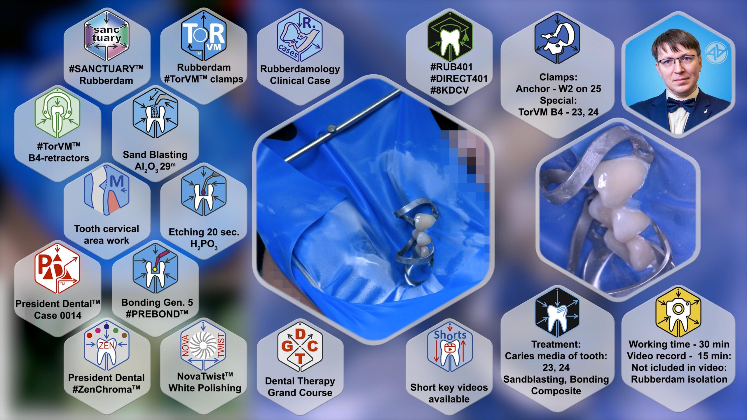
Every clinical case in this curriculum is analyzed through a custom-designed, 10-parameter data visualization interface. This interface surrounds the central live video stream, mapping the exact operational workflow of the procedure across the following distinct parameters:
- Rubber Dam Isolation Protocol: Identification of specific rubber dam plates and clamp selections (including standard anchors, Brinker series, or specialized soft-tissue retractors).
- Micro-Abrasive Surface Preparation: Specification of sandblasting parameters, explicitly detailing aluminum oxide ($Al_2O_3$) particle sizes utilized for biomimetic substrate conditioning.
- Acid-Etching Parameters: Precise timing protocols for selective or total-etch techniques on dentin and enamel.
- Adhesive System Execution: Detailed application time and protocol data for the bonding agent (e.g., Pre Bond, President Dental).
- Composite Selection & Stratification: Specification of the composite sub-types, viscosity, and precise shade mapping used to mimic natural tooth structure.
- Lining & Base Layering: Protocols for stress-decreasing steps, such as the implementation of bulk-fill materials or flowable liners in deep cavities.
- Clinical Case Indexing: A systematic cataloging number (e.g., Case 01) to cross-reference President Dental cases within the broader curriculum.
- Soft-Tissue and Contact-Point Management: Documentation of physical matrices, separation rings, and cervical retraction instruments (e.g., gingival retractors) required for anatomically correct proximal contours.
- Curriculum Mapping & Hyper-Targeted Navigation: Strategic indexing utilizing specialized identifiers (
#direct, specific chapter markers) and the#8KDCVquality standard. - Operational Metrics & Chronology: Real-time tracking of total chair time versus raw recorded video time, including transparent clinical callouts for omitted phases (e.g., instances where isolation or final polishing were modified due to patient-specific constraints).
3. The Tri-Camera Documentation Setup (4KDCV Standard)
To ensure absolute transparency and maximize the educational impact of each procedure, all clinical cases are documented using a synchronized, three-camera recording array formatted to the 4KDCV (4K Digital Clinical Video) standard. This setup eliminates visual blind spots and captures the micro-dynamics of material manipulation:
+-------------------------------------------------------------------+
|                           TOP CAMERA                              |
|             [Instrument Table / Tray Organization]                |
+-------------------------------------------------------------------+
         |                                                 |
         v                                                 v
+-----------------------------------+     +---------------------------------+
|        MAIN MACRO CAMERA          |     |          LATERAL CAMERA         |
|     [Microscopic Active Field]    |     |   [Operator Hands / Ergonomics] |
+-----------------------------------+     +---------------------------------+
- The Main Macro Camera (Microscopic Field): Positioned at the optical center, acting as a high-magnification clinical microscope. It captures the precise interaction between hand instruments, rotary burs, adhesive layers, and dental substrates.
- The Lateral/Face Camera: Positioned to the left, capturing the operator’s hands, direct instrument delivery, material handling, and the real-time stability of the rubber dam field.
- The Top/Overhead Camera: Positioned directly above the workspace to document the instrument tray layout, the status of materials, curing lamp placement, and overall workflow ergonomics.
4. Clinical Significance in Cariology
By combining multi-angle 4K video capture with an unambiguous 10-parameter clinical matrix, this methodology bridges the gap between theoretical cariology protocols and real-world clinical execution. It provides students and practicing dentists with a reproducible blueprint for everyday biomimetic restorations.
The clinical validation of any biomimetic restorative protocol relies on its predictability across diverse structural defects. Free-hand artistry cannot substitute for systematic engineering. Every cavity configuration introduces distinct stress distribution patterns, compliance values, and geometric boundaries that dictate how polymeric biomaterials behave under polymerization shrinkage and functional occlusal loads.
This chapter compiles the multi-format clinical registry of the #RSBC Protocol (Rubberdam, Sandblasting, Bonding, Composite) utilizing President Dental material systems. By analyzing these thirty clinical case portfolios—ranging from localized cervical erosion lines to total pre-endodontic coronal wall reconstructions—the clinician can master the integration of chemical, mechanical, and isolation strategies needed to ensure long-term restorative survival.
Comprehensive Reference Index (ADA Format)
- Anusavice KJ, Shen C, Rawls HR. Phillips’ Science of Dental Materials. 12th ed. St. Louis, MO: Elsevier Saunders; 2012.
- Broome JC, Ferracane JL, Hilton TJ. Summitt’s Fundamentals of Operative Dentistry: A Contemporary Approach. 4th ed. Hanover Park, IL: Quintessence Publishing; 2013.
- De Munck J, Van Landuyt K, Peumans M, Poitevin A, Lambrechts P, Braem M, Van Meerbeek B. A critical review of the durability of adhesion to tooth tissue: Methods and results. Journal of Dental Research. 2005;84(2):118-132.
- Ilie N, Hickel R. Resin composite restorative materials. Australian Dental Journal. 2011;56(1):59-66.
- Van Meerbeek B, Yoshihara K, Van Landuyt K, Vanherle G, Nor JE. From daily happiness to long-term durability in adhesion to tooth tissue. Japanese Dental Science Review. 2020;56(1):100126.
- Ferracane JL. Resin composite — State of the art. Dental Materials. 2011;27(1):29-38.
- Loomans BAC, Opdam NJM, Roeters FJM, Bronkhorst EM, Plasschaert AJM. Comparison of proximal contacts of Class II resin composite restorations in vivo. Operative Dentistry. 2006;31(6):688-693.
- Wirsching SM, Loomans BA, Hickel R. Proximal contact tightness and marginal integrity of posterior composite resin restorations under dynamic cyclic loading. Caries Research. 2023;57(2):142-151.
- Baba NZ. Contemporary Restoration of Endodontically Treated Teeth: Evidence-Based Diagnosis and Treatment Planning. St. Louis, MO: Springer Nature; 2018.
- Ritter AV, Boushell LW, Walter R. Sturdevant’s Art and Science of Operative Dentistry. 7th ed. St. Louis, MO: Elsevier; 2019.